

Free radicals in beer production impact flavor, color, and shelf life. Managing them is crucial for ensuring consistent brewing quality.
EPR Imaging and Spectroscopy
Novilet's innovative devices enhance the precision and sensitivity of EPR measurements, supporting groundbreaking research across diverse scientific fields.
EPR Imaging and Spectroscopy
Novilet's innovative devices enhance the precision and sensitivity of EPR measurements, supporting groundbreaking research across diverse scientific fields.
Discover Full Potential of EPR Imaging
Electron Resonance Imaging (ERI) allows for measuring materials that contain unpaired electrons and tracking the free radicals. The technique can be applied in many research projects in the areas of oncology research, neurodegeneration, inflammation and many more. Due to the fact that it gives the possibility to receive dynamic measurements and generate whole-body images, it may provide information about many unique microenvironments parameters and represent how they change in time.
Tracking the free radicals mechanisms can help to understand the origins and progressions of various diseases like cancer or neurodegenerative disorder. Redox state, antioxidant capacity or oxidative stress. These biophysical parameters play crucial roles in plenty of physiological mechanisms and pathological dysfunctions.
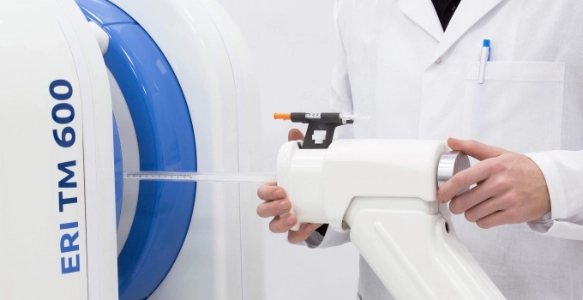
High-tech Solutions for EPR Spectroscopy
We recently designed the L-band spectrometer that is a perfect choice to carry out research on big objects, volumes and in-vivo samples. The spectrometer is equipped with a surface coil resonator that collects good-quality signals from precise localisation. With this device, researchers can study materials containing high water concentrations or perform research on mice, rats or humans(melanoma changes).
The eSPECT+ multiharmonic analyser is an expansion module that can be connected to X-Band Bruker Spectrometers. eSPECT+ increases the sensitivity of the spectrometer’s detection system even up to 10 times. It is a perfect solution for researchers struggling with weak signals. The expansion module allows for lowering the power level and researching live organisms. The eSPECT+ provides unlimited research possibilities and a new approach to classic EPR spectroscopy measurements. For more details, please contact a local Bruker representative.
Our Products
ERI Duo System

L-Band Spectrometer

eSpect+

Unlimited Applications
You may think that the technique can be applied mostly to in-vivo studies, but that is simply just the tip of the iceberg. Due to the fact that the method allows tracking of radicals’ activity and measuring big volumes and lossy samples, it has a huge potential in food or material science research.
Preclinical Research
Food Research
Material Science
Pharmaceutical Research
New technique
Endless possibilities
Recently Posted

Discover EPR research trends: in vivo applications, imaging, quantum science, machine learning, and spin probes transforming scientific advancements.

Interpreting Electron Paramagnetic Resonance (EPR) data requires understanding spectra, which reveal magnetic properties, structure, and dynamics of paramagnetic species. EPR spectra contain
Imaging or Spectroscopy? You Decide!
We know that having all the available options to explore is extremely important to the researchers. We provide solutions to perform both Imaging and Spectroscopy. In the imaging mode, the researcher can generate whole-body images that illustrate the measured microenvironment parameters. To collect the signal with even greater sensitivity the researcher can choose the surface coil resonator to measure the signal locally.